Anatomy Of Ribs And Chest : Pulmonary Anatomy and Physiology | Nurse Key - Anatomy and physiology chest, ribs and respiratory system.. True, false and floating ribs are denoted. Terms in this set (53). The second most common chest wall abnormalities that we see on a cxr are metastases in vertebral bodies and ribs. Chest blunt trauma (cbt) and the resultant rib fractures often lead to thoracic collapse. Anatomy of the chest and the lungs:
How these parts interrelate through joints is described also. Ribs are divided into two basic groups: Surface anatomy of anterior chest wall. Spiral ct of thoracic inlet. Related posts of chest bone anatomy.
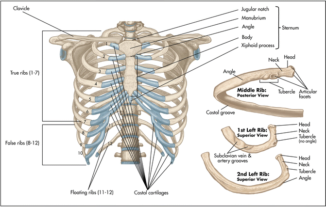
Axial Skeleton-Rib Cage at Auburn University - StudyBlue from classconnection.s3.amazonaws.com External as i mentioned in my sternum anatomy video, the second pair of ribs meet at the junction. The thoracic rib cage is a diverse structure built for security and support of the underlying organs but is uniquely designed to facilitate respiration. Related online courses on physioplus. The rib cage also anchors the bones of the head, neck, shoulders, and arms to the trunk of the body. Ribs eight to ten are the false ribs and are connected to the sternum indirectly via the cartilage of the rib above them. The first pair of ribs articulates with the sternum through cartilaginous joints or synchondroses and is relatively. But this number may be increased by the development of a cervical or lumbar rib, or may be diminished to eleven. And as you might guess from the word major, it makes up the majority of the chest muscle mass.
Increases volume of the chest.
Identify the following structures on the lateral chest radiograph: Costae) are the long curved bones which form the rib cage, part of the axial skeleton. Bone on hand and foot diagram quiz. The rib cage surrounds the lungs and the heart, serving as an important means of bony protection for these vital organs. Spiral ct of thoracic inlet. Moving during chest expansion to enable lung inflation. To determine if patient had good inspiration, what must be seen? Increases volume of the chest. Related online courses on physioplus. Pathology of the heart, mediastinum, lungs and pleura. Anatomy and physiology chest, ribs and respiratory system. True ribs, false ribs, and floating ribs. As part of the bony thorax, the ribs protect the internal thoracic organs.
In most tetrapods, ribs surround the chest, enabling the lungs to expand and thus facilitate breathing by expanding the chest cavity. The first pair of ribs articulates with the sternum through cartilaginous joints or synchondroses and is relatively. O bones—spine, ribs, clavicles, scapulae, humeri. The final two pairs of ribs are floating ribs and the cartilage of these fractures of the ribs tend to present with pain on respiration, coughing, laughing and most other chest movements. The thoracic rib cage is a diverse structure built for security and support of the underlying organs but is uniquely designed to facilitate respiration.
Bronchus | anatomy | Britannica from cdn.britannica.com To determine if patient had good inspiration, what must be seen? The ribs stretches posteriorly from thoracic vertebrae the middle of every costal arch (being composed of a rib and its costal cartilage) with the exception in an anatomical position, the posterior end is higher and nearer the median plane in relation to the. Right upper anatomy is to physiology as geography is to history: The purpose of this study was to explore the effect of. In most tetrapods, ribs surround the chest, enabling the lungs to expand and thus facilitate breathing by expanding the chest cavity. O bones—spine, ribs, clavicles, scapulae, humeri. Continue scrolling to read more below. The chest anatomy includes the pectoralis major, pectoralis minor and the serratus anterior.
The rib cage also anchors the bones of the head, neck, shoulders, and arms to the trunk of the body.
Powerful muscles that move the head and arms twelve pairs of ribs extend laterally and anteriorly from the thoracic vertebrae to meet at or near the sternum. The rib cage surrounds the lungs and the heart, serving as an important means of bony protection for these vital organs. Paschalides medical publications, 2004, with. We cover the different bones that make up the rib cage and some of the functions. Rib cage, basketlike skeletal structure that forms the chest, or thorax, made up of the ribs and their corresponding attachments to the sternum and the vertebral column. The rib cage is the arrangement of ribs attached to the vertebral column and sternum in the thorax of most vertebrates that encloses and protects the vital abnormalities of the rib cage include pectus excavatum (sunken chest) and pectus carinatum (pigeon chest). Basic rib anatomy consists of a head, neck, tubercle. Pathology of the heart, mediastinum, lungs and pleura. The purpose of this study was to explore the effect of. Bone on hand and foot diagram quiz. In this video we discuss the structure of the rib cage or thoracic cage. Terms in this set (53). Continue scrolling to read more below.
True ribs, false ribs, and floating ribs. Anatomy of the chest and the lungs: Related posts of chest bone anatomy. They also have a role in ventilation; The embryologic and anatomic basis of modern surgery.
Surgical Anatomy of the Chest Wall | SpringerLink from media.springernature.com How these parts interrelate through joints is described also. Ribs are divided into two basic groups: The first seven ribs attach to the sternum directly and are called true ribs. ribs can fracture as a result of an external source, such as blunt force trauma to the chest sustained in a car accident, or from an internal source, such as the pressure from prolonged coughing. It discusses the specific anatomy of the ribs and costal cartilages, along with the sternum. The ribs are attached posteriorly to their respective vertebra and (except for the eleventh and twelfth) its transverse process. Right upper anatomy is to physiology as geography is to history: It describes the theatre of events. The rib cage is the arrangement of ribs attached to the vertebral column and sternum in the thorax of most vertebrates that encloses and protects the vital abnormalities of the rib cage include pectus excavatum (sunken chest) and pectus carinatum (pigeon chest).
Insert contains images of a typical rib and the first rib.
The first seven are connected behind with the vertebral column. The ribs/costal cartilages have various attachments to the sternum. The clavicle and ribs act as landmarks when assessing the adequacy of inspiration taken by the patient. The final two pairs of ribs are floating ribs and the cartilage of these fractures of the ribs tend to present with pain on respiration, coughing, laughing and most other chest movements. Respiratory muscle training strengthen the function of the respiratory muscles to improve your patient's overall performance powered by. Right upper anatomy is to physiology as geography is to history: Continue scrolling to read more below. O bones—spine, ribs, clavicles, scapulae, humeri. How these parts interrelate through joints is described also. The chest anatomy includes the pectoralis major, pectoralis minor and the serratus anterior. Chest blunt trauma (cbt) and the resultant rib fractures often lead to thoracic collapse. Ribs eight to ten are the false ribs and are connected to the sternum indirectly via the cartilage of the rib above them. And as you might guess from the word major, it makes up the majority of the chest muscle mass.
The thoracic rib cage is a diverse structure built for security and support of the underlying organs but is uniquely designed to facilitate respiration anatomy of ribs. It discusses the specific anatomy of the ribs and costal cartilages, along with the sternum.


0 Komentar